GenEgoista
Baneado
- Desde
- 4 Feb 2013
- Mensajes
- 3.566
- Reputación
- 3.412
Tema de hoy: El magufo mayor del blog TIRA LA TOALLA con el tema VIH/SIDA ante los OWNEDS que le hemos hecho comer en este foro.
Eso sí, la toalla envuelve un pedrusco que pretende hacer daño pero es de pura plastilina.
---------- Post added 07-may-2013 at 13:40 ----------
Eso sí, la toalla envuelve un pedrusco que pretende hacer daño pero es de pura plastilina.
Desmontemos estas afirmaciones NO REFERENCIADAS una por una y con las referencias correspondientes:¿Y a mí que más me da?: una despedida personal del “negacionismo” del SIDA
"...Había pensado arrancar el artículo explicando todas las pruebas realizadas con las muestras de esos tubos que llevan a la conclusión de que el contenido del mismo es un bichito infeccioso que se ha denominado VIH: (1) el contenido de la muestra mata linfocitos, (2) la observación al microscopio electrónico permite observar la morfología típica del bichito, (3) presenta actividad retrotranscriptasa, (4) en ratones “humanizados” produce la muerte de sus linfocitos T humanos, (5) debe trabajarse en condiciones de precaución y en nivel de bioseguridad 3 porque se han producido infecciones accidentales en su manipulación, (6) la secuenciación de su genoma muestra la elevada homología con los lentivirus, una variedad de retrovirus, y no tienen homología con secuencias humanas. Eso por no hablar de que su modificación genética permite desarrollar herramientas de terapia génica o estudiar “in vitro” la dinámica de la infección..."
(1) el contenido de la muestra mata linfocitos,
Los retrovirus han sido objeto de estudio en la lucha contra el cáncer precisamente por no ser citocidas (no dar de baja de la suscripción de la vida a la célula huésped).
En el caso del "VIH", las proteinas usadas en las pruebas diagnósticas se producen en masa mediante cultivo en líneas de linfocitos inmortales como la H9, que no sufren daño alguno a pesar de estar supuestamente infectadas con VIH:
"...Gallo retrovirologists Mikulas Popovic had grown it (HIV) in large quantities for the first time, in cells he dubbed H9 that were not killed by the bichito..."
The untold story of HUT78

Los linfocitos supuestamente "infectados" con "VIH" se replican normalmente, de forma "crónica" (es decir, no mueren) y son los medicamentos, no el supuesto bichito, quienes acaban con ellas:
Inhibition of human immunodeficiency bichito and growth of infected T cells by the immunosuppressive drugs cyclosporin A and FK 506.
The untold story of HUT78
Los linfocitos supuestamente "infectados" con "VIH" se replican normalmente, de forma "crónica" (es decir, no mueren) y son los medicamentos, no el supuesto bichito, quienes acaban con ellas:
Inhibition of human immunodeficiency bichito and growth of infected T cells by the immunosuppressive drugs cyclosporin A and FK 506.
targets for anti-HIV agents.
MATERIALS AND METHODS
Assay for the Effects of the Drugs on HIV Cell Fusion and
Replication.
We have used a clone of the leukemia T-cell line
Molt 4 that expresses a very high level of CD4. This was
infected with the highly cytopathic NDK strain of HIV-1 (15)
and cells that are chronically infected with the bichito but that
replicate readily were selected. When these HIV-infected
Molt 4 cells were cocultivated with uninfected Molt 4 cells at
a ratio of 100 noninfected:1 infected cell, very large giant cells
were formed within 24 hr (Fig. 1). We selected, in the same
manner, cells chronically infected with the Cambridge isolate
of HIV-2 (16). The formation of giant cells within 24 hr was
used in the mixed culture as a rapid assay to determine the
anti-HIV effect of CsA and FK 506. In addition, we have
tested the cytotoxicity and anti-HIV-1 activity of these
substances in an anti-HIV assay system that has been described
previously (17). In this assay system, target cells were
preincubated overnight with the compound being tested
before infection at a multiplicity of 1 infectious unit of HIV-1
---------- Post added 07-may-2013 at 13:40 ----------
(2) la observación al microscopio electrónico permite observar la morfología típica del bichito,
La morfología "típica del bichito" lo es también de los exosomas que son partículas mensajeras de origen celular:
por tanto, los exosomas humanos es lo que estos FARSANTES están revendiendo como "un nuevo bichito mortífero":
Conclusión lógica: LAS PARTICULAS DE "VIH" NO MAS QUE SIMNPLES EXOSOMAS.
Un reciente estudio del 2008 demuestra que las proteinas GAG del "VIH" proceden EN REALIDAD de los exosomas:
Esto invalida todas las pruebas del VIH que detectan anticuerpos a las proteinas GAG, entre ellos el Western Blot confirmatorio.
Por si esto fuera para demostrar que nos venden exosomas al precio de VIH, los supuestos "aislamientos del VIH" están groseramente contaminados por:
Por útimo, partículas idénticas al "VIH" han sido detectadas en pacientes NO INFECTADOS:
La morfología "típica del bichito" lo es también de los exosomas que son partículas mensajeras de origen celular:
- con la misma topología que el "VIH",
- de la misma talla que el "VIH",
- con las mismas moléculas que el "VIH"
- de la misma talla que el "VIH",
- con las mismas moléculas que el "VIH"
por tanto, los exosomas humanos es lo que estos FARSANTES están revendiendo como "un nuevo bichito mortífero":
Higher-Order Oligomerization Targets Plasma Membrane Proteins and HIV Gag to Exosomes
"human immunodeficiency bichito (HIV) particles bud from these two cell types at the same sites as exosomes, have the same topology as exosomes, have a similar size as exosomes, and are enriched in the same molecules as exosomes"
"human immunodeficiency bichito (HIV) particles bud from these two cell types at the same sites as exosomes, have the same topology as exosomes, have a similar size as exosomes, and are enriched in the same molecules as exosomes"
Conclusión lógica: LAS PARTICULAS DE "VIH" NO MAS QUE SIMNPLES EXOSOMAS.
Un reciente estudio del 2008 demuestra que las proteinas GAG del "VIH" proceden EN REALIDAD de los exosomas:

Esto invalida todas las pruebas del VIH que detectan anticuerpos a las proteinas GAG, entre ellos el Western Blot confirmatorio.
Por si esto fuera para demostrar que nos venden exosomas al precio de VIH, los supuestos "aislamientos del VIH" están groseramente contaminados por:
1. exosomas.
Ya vimos que la proteina GAG p24 es característica de los exosomas, y a pesar de este conocimiento se usa rutinariamente para diagnosticar VIH y SIDA en personas sanas (pruebas ELISA y Western Blot).
Los exosomas forman parte del propio sistema celular, son partículas mensajeras que transmiten RNA entre células. Habría que ver cuantos "bichito" de los atlas médicos son en realidad exosomas celulares
2. vesículas celulares
Discrimination between exosomes and HIV-1:... [J Immunol Methods. 2008] - PubMed - NCBI
In this study, HIV-1 particles or exosomes in cell-free supernatant filtered through 0.22-µm membrane were concentrated by ultracentrifugation at 100,000 ×g for 45 min, re-suspended and then separated by centrifugation through a 6–18% Optiprep™ velocity separation gradient. Dettenhofer et al. was the first to describe efficient separation of microvesicle contaminants from HIV-1 preparations (Dettenhofer and Yu, 1999). This efficiency is confirmed in Fig. 1, which shows p24gag protein concentrated mainly in fraction 15.6.
In this study, HIV-1 particles or exosomes in cell-free supernatant filtered through 0.22-µm membrane were concentrated by ultracentrifugation at 100,000 ×g for 45 min, re-suspended and then separated by centrifugation through a 6–18% Optiprep™ velocity separation gradient. Dettenhofer et al. was the first to describe efficient separation of microvesicle contaminants from HIV-1 preparations (Dettenhofer and Yu, 1999). This efficiency is confirmed in Fig. 1, which shows p24gag protein concentrated mainly in fraction 15.6.
Ya vimos que la proteina GAG p24 es característica de los exosomas, y a pesar de este conocimiento se usa rutinariamente para diagnosticar VIH y SIDA en personas sanas (pruebas ELISA y Western Blot).
Los exosomas forman parte del propio sistema celular, son partículas mensajeras que transmiten RNA entre células. Habría que ver cuantos "bichito" de los atlas médicos son en realidad exosomas celulares
2. vesículas celulares
Cell Membrane Vesicles Are a Major Contaminant of Gradient-Enriched Human Immunodeficiency bichito Type-1 Preparations
".. Electron microscopy of gradient-enriched preparations from supernatants of bichito-infected cells revealed an excess of vesicles with a size range of about 50 – 500 nm, as opposed to a minor population of bichito particles of about 100 nm..."
"...We have analyzed gradient-enriched bichito preparations and found that there is contamination with an excess of nonviral membrane vesicles of cellular origin. This was the case when the bichito was grown in an immortalized cell line or in activated peripheral blood mononuclear cells (PBMC). These vesicles, which are released from both noninfected and HIV-1 infected cells, contain a selection of cellular membrane proteins similar to, but not identical to those in the bichito particles. ..."
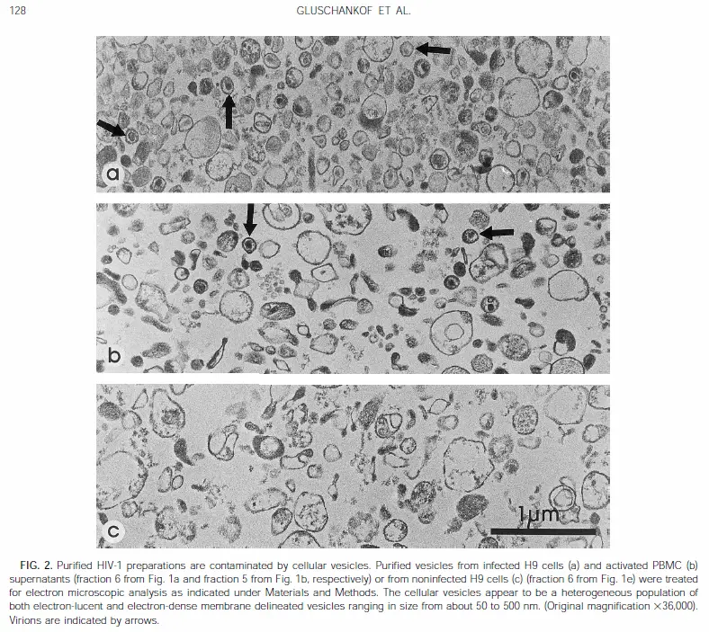
Por útimo, partículas idénticas al "VIH" han sido detectadas en pacientes NO INFECTADOS:
Ante todo la referencia indexada en Pubmed:
Hum Pathol. 1988 May;19(5):545-9.
The ultrastructural and immunohistochemical demonstration of viral particles in lymph nodes from human immunodeficiency bichito-related and non-human immunodeficiency bichito-related lymphadenopathy syndromes.
O'Hara CJ, Groopman JE, Federman M.
Source
Department of Pathology, England Deaconess Hospital, Harvard Medical School, Boston, MA 02215.
Abstract
Viral particles have been demonstrated by electron microscopy in lymph nodes from patients with acquired immune deficiency syndrome AIDS-related persistent generalized lymphadenopathy (PGL) syndrome. Immunohistochemical and in situ hybridization studies have identified these viruses as the human immunodeficiency bichito (HIV). In this study, we examined 20 PGL lymph nodes and found viral particles in 18 cases. Immunohistochemical studies on these cases revealed positive staining for the HIV core protein P24 within germinal centers of secondary follicles. In addition we found viral particles, morphologically indistinguishable from those observed in PGL lymph nodes, in 13 of 15 non-HIV related reactive lymph nodes. Immunohistochemical staining of these lymph nodes for the P24 core protein was negative. None of the patients in this group had risk factors for developing AIDS and none exhibited clinical evidence of immune deficiency. We conclude that the viral particles observed in PGL lymph nodes are most likely HIV, but similar particles can be seen in reactive lymph nodes not associated with HIV infection. The discrete localization of these particles within germinal centers has been observed for other viruses and immune complexes and a possible mechanism of this antigen deposition is discussed.
The ultrastructural and immunohistochemical demon... [Hum Pathol. 1988] - PubMed - NCBI
Les traduzco lo subrayado: Encontraron "partículas indistinguibles" del "VIH"... en pacientes SIN "VIH" :8:.
Cuando me encontré con este abstract increíble, a la primera oportunidad oportunidad que tuve corrí a mi Alma Mater a leer el artículo completo. Sí: el artículo respalda exactamente lo que dice el Abstract. Aún me recuerdo tembloroso leyendo el polvoriento tomo de "Human Pathology" en el mismo archivo de mi Universidad. Ni siquiera me senté para leerlo. Allí estaba la Piedra Filosofal de todo disidente del SIDA: El artículo que demostraba que había "VIH"...en muestras "cocultivadas" de NO "INFECTADOS" con "VIH".
El Grupo de Perth ya había notado la existencia de este extraordinario artículo, y habían dedicado comentario al escaneado de las fotos de este artículo:
The Perth Group HIV-AIDS Debate Website
En el punto 4 el Grupo de Perth enlaza las micrografías de O'Hara 1988

Aquí "VIH" en paciente "infectado por el VIH".

Aquí, partícula "morfológicamente indistinguible del VIH" en persona... sin "VIH"....
Última edición: